


Acta Microscopica
REVIEW: HYDRO-, MICRO- AND NANOGELS STUDIED BY COMPLEMENTARY MEASUREMENTS BASED ON SEM AND SFM.
Matzelle, T.*, Reichelt, R.
*Chimie-Physique des Matériaux, Université Libre de Bruxelles, B-1050 Bruxelles, Belgium
Institut für Medizinische Physik und Biophysik, Universitätsklinikum, Westfälische Wilhelms-Universität, Münster, Germany
*Corresponding author: matzelle@ulb.ac.be
Received: March 11th, 2008. Accepted: May 2nd, 2008
Published on-line: May 30, 2008
ABSTRACT
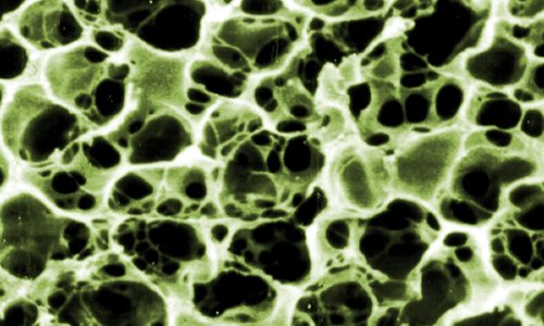
The research on soft materials is interdisciplinary. In the present work we focus on “smart hydrogels” as most promising representatives studied by complementary Scanning Force Microscopy (SFM) and high resolution Scanning Electron Microscopy (SEM). The extremely large range of water uptake of these hydrogels (up to 103 times of their mass) on one hand and their distinct softness (Young´s modulus < some tens of kPa) on the other hand constitute real challenges for the characterization of their local structure, the caging of nano-scaled particles, and some micromechanical properties. SFM images were compared with those obtained by SEM in the dry, swollen and deswollen state of the “smart gel” PNIPAAm [poly-(N-isopropylacrylamide)]. PNIPAAm reacts on tiny variations of the temperature around 33°C by significantly changing its volume. While both imaging techniques revealed very similar results concerning the surface structure in the dry state, highly resolved structural features remained unapproachable for SFM in the wet state. In a contrary, SEM at high resolution revealed after state-of-the-art cryo-preparation, freeze-drying and subsequent ultrathin Pt/C-coating a sponge-like structure with cavity sizes of around 40 nm in the swollen state and 20 nm in the deswollen state. SFM proved to be an appropriate instrument revealing the local elastic surface properties. For example, at the swollen state at 10 °C, the Young’s modulus was found to be more than 100 times lower than for the deswollen state at 35 °C. In addition, SEM also proved to be very suitable for the structural research of microgels and filled hydrogels. The application of both SFM and SEM contribute complimentarily to a characterization of different hydrogel systems at different states.
RESUMEN
La investigación en materiales blandos es interdisciplinaria. En este trabajo nos concentraremos en “hidrogeles smart” como representantes más prometedores estudiados complementariamente por Microscopía de Fuerza Atómica (MFA) y Microscopía Electrónica de Barrido (MEB) de alta resolución. Los hidrogeles muestran una alta capacidad de absorción de agua (hasta 103 veces su masa) y un amplio rango de propiedades mecánicas (Módulo de Young < algunas decenas de KPa). El estudio sistemático de estas propiedades y su estrecha relación con la microestructura constituyen un verdadero desafío para la caracterización de su estructura local, la incorporación y distribución de nanopartículas en su estructura interna y algunas propiedades micromecánicas. En esta revisión se hace un análisis comparativo de imágenes obtenidas desde SFM y SEM del “smart gel” PNIPAAm [poli-(N-isopropil-acrilamida)] en estado xerogel, hinchado y deshinchado. El PNIPAAm modifica significativamente su volumen ante pequeñas variaciones de temperatura alrededor de 33 ºC. Mientras ambas técnicas revelaron resultados muy similares relativos a la estructura superficial en el estado seco, las características estructurales de mayor resolución han permanecido inalcanzables para el SFM en estado húmedo. Al contrario, el SEM a alta resolución después de una crio-preparación, congelación-secado y subsiguienre deposición de una capa ultrafina de Pt/C, revela una estructura esponjosa con cavidades de dimensiones cercanas a los 40 nm en su estado hinchado y 20 nm en el estado deshinchado. SFM ha demostrado ser un instrumento apropiado para revelar las propiedades locales elásticas superficiales. Por ejemplo, en el estado hinchado a 10 ºC, el módulo de Young era más de 100 veces el del estado deshinchado a 35 ºC. Adicionalmente, SEM ha demostrado ser muy adecuado para la investigación estructural de microgeles e hidrogeles nanoestructurados. La aplicación de SFM y SEM contribuyen complementariamente a la caracterización de diversos sistemas de hidrogeles en diferentes estados.
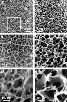
FESEM secondary electron micrographs of the PNIPAAm gel surface in the deswollen state at 35 °C (a - c) and the swollen gel surface at room temperature (d - f) at different magnifications. Both states show a similar spongy structure but the mean size of cavities differs by a factor of approximately two. Reprinted with permission from J Phys Chem B 106:2861-2866, Copyright (2002) ACS.
Keywords
SFM, SEM, hydrogels, microgels, Young´s modulus